Introduction to breathing assessment
Breathing assessment is essential in determining an individual’s respiratory function. One of the fundamental aspects of patient evaluation, breathing assessment provides valuable insights into pulmonary function and informs clinical management decisions. Understanding the components of this assessment is crucial in ensuring accurate diagnosis and treatment plans.
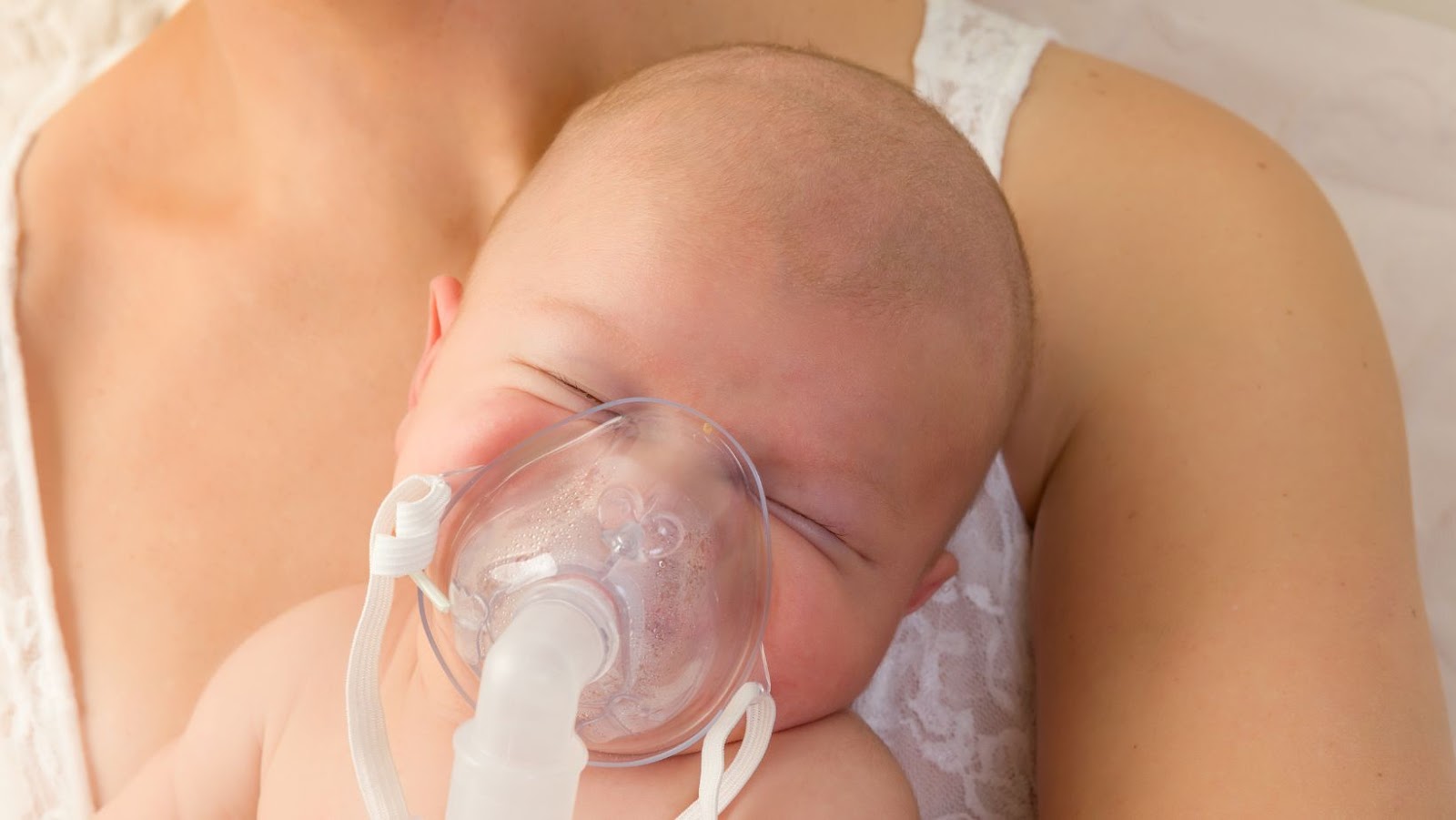
In conducting a breathing assessment, one key component is evaluating respiration rate and effort. This involves monitoring how quickly a patient breathes, along with difficulty or discomfort associated with breathing. Another important aspect to consider is lung sounds, where clinicians listen for abnormal breath sounds that could indicate underlying respiratory pathologies. Additionally, oxygen saturation levels must be measured using pulse oximetry.
It is noteworthy that chest expansion examination through palpation can also provide useful information about respiratory health. To gain more insight into respiratory function, spirometry can be performed to measure airflow volumes during inhalation and exhalation completely.
As part of a routine medical check-up, John visited his physician for a breathing assessment. The doctor first conducted an initial examination by checking John’s pulse rate, blood pressure and listened to heart sounds followed by a detailed examination of his lungs using a stethoscope to assess any abnormal lung sounds or murmurs noted before concluding the test with spirometry examination that revealed he had mild asthma requiring bronchodilator therapy during exacerbations.
Breathing assessment is so important, even Darth Vader would approve.
Importance of breathing assessment
Maintaining healthy lungs is crucial for a cancer patient’s overall well-being. Assessing breathing helps detect the early signs of diminished lung function, allowing suitable remedies to be prescribed promptly. Evaluating the quality and quantity of air ventilation, and identifying abnormalities in lung sounds are integral components of this assessment.
The observation and measurement of proper breathing techniques can provide various insights into an individual’s overall health status. Not only does it help diagnose respiratory illnesses, but it also helps evaluate any underlying conditions. Abnormalities in breath sounds, such as wheezing or crackling, could indicate infections or inflammations in the lungs, whereas shallow breathing can result from chronic obstructive pulmonary disease (COPD). Examining breathing patterns during physical exercise is also crucial in evaluating aerobic endurance levels.
Finally, during these assessments, healthcare professionals need to communicate effectively with patients by explaining the purpose and procedure for any tests performed. Patients should be given guidance on the importance of deep breaths for their recovery and how practicing different breathing techniques can enhance their lung function.
Pro Tip: Encourage patients to practice regular deep-breathing exercises to improve lung capacity and promote relaxation. Take a deep breath and relax, because if you’re not prepared for the assessment, you might just end up hyperventilating.
Preparing for the assessment
To prepare for your breathing assessment with its various components, including informed consent, hygiene protocols, and an explanation of the procedure, you need to take a few steps. You should be informed about the process, understand the hygiene protocols that need to be followed, and give consent before the assessment begins. Understanding these sub-sections can help you ensure that you have a smooth and successful breathing assessment.
Informed consent
Before proceeding with the assessment, it is important to ensure that participants have provided Validated Informed Consent. This means that each participant has received sufficient information about the study and its requirements to make an informed decision about whether or not they want to participate. It also includes ensuring that individuals understand any risks and benefits associated with participating in the study, as well as their right to withdraw at any time.
To obtain Validated Informed Consent, researchers should provide participants with a clear description of the study’s purpose, methods, procedures, and anticipated results. Researchers should also offer explanations of any potential risks or discomforts involved in the study. Additionally, researchers must offer assurances of confidentiality measures for participant data.
It is essential to use appropriate language while obtaining Informed Consent by avoiding any confusing terminology or jargon. Questions should be answered appropriately so that participants can make an informed decision based on accurate information.
Undeniably, research conducted without Properly Obtained Validated Informed Consent can be classified as unethical behavior within a scientific community. Therefore it is necessary always to gain Validated Informed Consent from all participants before conducting any research.
Remember, good hygiene isn’t just important for passing your assessment, it’s also essential for not being gross.
Hygiene protocols
To ensure compliance with hygienic requirements, consider implementing sanitation procedures. Use disposable gloves, masks, and hairnets while preparing or handling food items. Check the expiration dates of all products before using them. Keep the surfaces clean by disinfecting countertops, equipment and refrigerator on a regular basis.
Also, segregate cooked food from raw food to prevent cross-contamination. In addition, make it mandatory for all staff members to wash their hands with soap frequently. Following these guidelines can help reduce the spread of harmful microorganisms.
Pro Tip: Maintaining high hygiene levels helps in preventing unwanted contamination and provides a safer environment for employees and customers alike.
Get ready to be poked, prodded, and tested like a lab rat – but don’t worry, it’s all in the name of assessment.
Explanation of the procedure
Preparing for an assessment is a crucial step towards achieving the desired outcome. To ensure the smooth running of the process, one needs to prepare adequately and understand what lies ahead.
Here is a simple and concise three-step guide to help you with your assessment preparation:
- Understand the scope of the assessment: The first step involves getting familiar with the assessment format and requirements. Gaining knowledge on these aspects will guide you in setting realistic goals, allocating sufficient time, and focusing on key areas that require attention.
- Organize your study materials: After understanding what is expected from you, gather your study materials such as textbooks, notes, and any other online resources available. Ensure to revise all relevant information on each concept that will be assessed.
- Practice mock assessments: The final step is conducting practice assessments to gauge how well prepared you are. This stage provides insights into where to improve before taking the actual assessment.
In addition, it’s essential to maintain a healthy mindset during preparations. Take enough breaks amid studies, exercise regularly, prioritize sleep, eat healthily and hydrate often.
It may interest you to note that research has shown individuals who actively engage in self-assessments go on to perform better than those who don’t – Schoendorfer et al., (2019).
Therefore, take this opportunity of preparing adequately as it increases chances of performing optimally in assessments.
Breathing assessment – because if you can’t breathe, nothing else matters… except maybe getting someone to do the assessment for you.
What are the components of the breathing assessment
To assess someone’s breathing accurately, observing their breathing, measuring their respiratory rate, auscultating their lung sounds, taking their pulse oximetry, analyzing their arterial blood gas and performing chest x-ray can determine how well their lungs are working. These sub-sections serve as solutions to the components of the breathing assessment, providing a comprehensive view of one’s respiratory health.
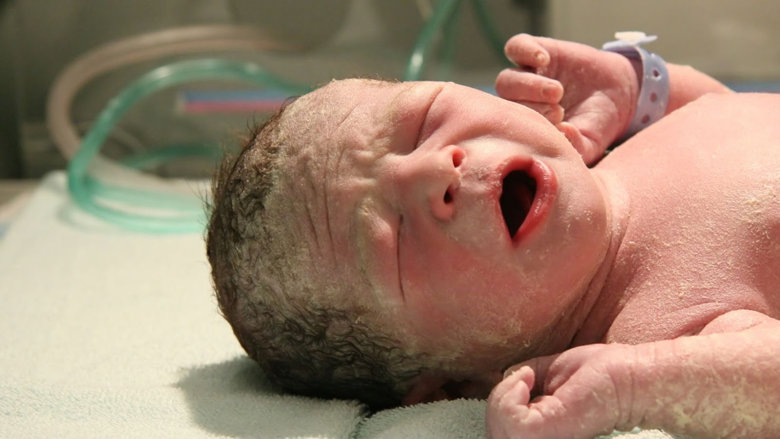
Observation of breathing
Assessment of respiration incorporates many components including the visual observation of breathing patterns. An astute observer can detect nuances in breathing such as rapid or shallow breaths, irregularity, or signs of distress. This component also includes the assessment of oxygen saturation levels via pulse oximetry and recording objective data on respiratory rate and depth. Recognizing subtle changes in breathing is vital for early recognition of patient deterioration.
The visual observation of breathing encompasses monitoring both the rate and depth at which an individual takes each breath. A clinician observes the chest movements, identifies any flaring at the nostrils, sucks in between or under ribs, abdominal movement with each breath and any sounds heard while breathing such as wheezes, stridor or crackles. The observations noted may suggest underlying conditions like asthma, bronchitis or pneumonia which has to be obtained through further evaluations if detected.
Correct respiratory assessments require clinicians to observe patients using a systematic approach regardless of their age or previous health conditions. Timely recognition of changes in normal respiratory patterns is crucial to prevent clinical deterioration that may result in negative health outcomes for patients. As clinicians assess this significant component during respiratory evaluations, they must remain alert and focused on identifying even slight deviations from typical breathing norms.
Inadequate assessments can lead to delayed diagnosis, potentially compromising patient outcomes; therefore thorough evaluation is paramount. Accurate medical assessments foster precision treatment plans culminating into high-quality patient care. By taking a systematic approach during evaluation helps medical practitioners appropriately identify abnormalities, take appropriate steps to address deteriorating symptoms before they advance further and re-evaluate treatment plans during follow-up visits encouraging quality healthcare practices using evidence-based interventions depending on findings; avoiding unwanted complications for patients.
It’s imperative clinicians incorporate assessment of respiration as part and parcel it with most clinical evaluations incorporating enhanced patient safety protocols aimed at improving healthcare delivery ensuring nothing goes unobserved for better patient outcomes overall. If you’re looking for an excuse to stare at someone’s chest without being creepy, just tell them you’re measuring their respiratory rate.
Measurement of respiratory rate
Respiratory rate is a crucial component to assess breathing patterns. The assessment involves measuring inhalation and exhalation frequency in units of breaths per minute. Accurate measurement of respiratory rate yields essential information on overall lung health, oxygen levels, carbon dioxide levels, and ventilation effectiveness. The process is simple and non-invasive, requiring minimal patient cooperation.
Measuring respiratory rates can be done through different methods such as visual observation or using electronic devices. While the visual method only offers an estimate of respiratory rate (RR), electronic methods offer greater accuracy in monitoring trends over prolonged periods. These devices include pulse oximeters, capnometers, and respirometers.
The quality of respiratory measurements can be affected by various factors such as age, emotional state, body position, medication use, lung disease presence among others. Medical practitioners must consider these critical variables when conducting assessments to get an accurate diagnosis and prescribe effective treatment plans.
Not evaluating or misinterpreting respiratory rates can lead medical practitioners to overlook vital signs relating to breathing patterns which may result in poorer patient outcomes. Accurate measurement of the respiratory rate is a matter of utmost importance in identifying early warning signs for conditions that require immediate intervention or emergency department attention leading to better health outcomes.
Auscultation of lung sounds
When it comes to assessing respiratory function, one crucial component is the evaluation of lung sounds using auscultation. This diagnostic technique involves listening to the sounds produced by the lungs during breathing.
During auscultation, a healthcare provider places a stethoscope on various areas of the chest, including the front and back. Using this approach, they can identify normal breath sounds and abnormal ones, such as wheezing or crackling.
One essential tool in auscultating lung sounds is understanding their characteristics. Normal breath sounds include vesicular, bronchovesicular, and bronchial breath sound patterns. Abnormalities may include wheezes from narrowed airways or crackles from fluid in the lungs.
Consequently, it becomes imperative that every healthcare provider knows how to assess lung sounds accurately and recognize potential issues promptly. Failure to grasp these skills could lead to misdiagnosis or missed diagnoses of respiratory disorders. Therefore, regular training can improve consistency and accuracy of evaluations.
Thus, health care providers should consistently seek professional development opportunities to ensure optimal patient outcomes.
Looks like we’ll be getting intimate with our fingers again as we explore the world of pulse oximetry.
Pulse oximetry
Measuring the oxygen saturation levels in the blood using a non-invasive procedure is an essential component of breathing assessment. This procedure is known as Oxygen Saturation Monitoring, and it involves using a device called Pulse Oximeter.
The oximeter comprises a probe-like sensor placed on the fingertips or other body parts that can accurately detect oxygen saturation levels. The machine uses two lights of different wavelengths to measure the amount of light absorbed by oxygenated and non-oxygenated blood. The measurements taken by pulse oximetry are reliable and convenient indicators of respiratory distress, hypoxemia, or heart diseases.
It’s important to know that factors like poor vascular perfusion, decreased cardiac output, cold temperature, or dark nail polish can affect the accuracy of pulse oximetry readings. Therefore, maintaining proper hand positioning and ensuring good contact between skin and sensor is critical for accurate results.
The use of pulse oximetry during breathing assessment allows for timely diagnosis and management of respiratory disorders. Healthcare professionals should be knowledgeable about its usage and limitations.
To ensure maximum accuracy when using pulse oximetry for measurement purposes, one should consider calibrating the machine regularly at specified intervals and recording observations while considering external influencing factors.
You know you’re in a medical profession when the words ‘arterial blood gas analysis‘ no longer scare you, they just give you a rush of excitement.
Arterial blood gas analysis
When assessing breathing, analyzing the oxygen and carbon dioxide levels in arterial blood is crucial. This test measures the partial pressure of oxygen, carbon dioxide, and pH levels in the arterial blood to determine if respiratory failure or hypoxia is present. Here is the normal range for each parameter in the arterial blood gas test:
| Parameter | Normal Range |
| pH | 7.35-7.45 |
| pCO2 (partial pressure of carbon dioxide) | 35-45 mm Hg |
| pO2 (partial pressure of oxygen) | 80-100 mm Hg (breathing room air at sea level) |
| Bicarbonate (HCO3-) | 22-28 mEq/L |
| Oxygen saturation (SaO2) | >90% Good tolerance for activity with SpO2 =<93% |
The arterial blood gas test helps in diagnosing acidity/alkalinity imbalances and respiratory or metabolic derangements. Low values indicate hypoxemia with relative hypocapnia, while high values can imply hypercapnic respiratory failure or compensated metabolic alkalosis. Seek medical attention early if symptoms persist, as untreated respiratory conditions can be life-threatening. If the chest x-ray doesn’t show any abnormalities, congratulations, your lungs are boring.
Chest x-ray
Assessing the respiratory system involves various components, one of which is a medical imaging technique that provides visual insights into the chest area. This technique allows clinicians to analyze and evaluate the lungs’ condition by capturing X-rays of the chest region via radiation exposure.
The X-ray can indicate any disruptions in lung functions such as inflammation, tumors, fluid buildup and identify potential lung conditions like pneumonia or lung cancer. The images generated are usually reviewed in conjunction with other tests and examinations to provide a holistic understanding of a patient’s health.
It’s important to note that while chest X-rays are widely used due to their effectiveness in evaluating the respiratory system, they’re not always able to identify underlying conditions, particularly those that manifest themselves only in later stages.
According to the American College of Radiology (ACR), its Collaborative Imaging Clinical Decision Support System may identify chest x-rays as sufficient when dealing with specific issues like pneumonia or assessing a patient for metasticesis; however CT scans carry more robust accuracy when diagnosing a range of pulmonary issues including subtle pathologies.
Therefore, it’s essential for healthcare professionals to integrate multiple diagnostic approaches and methods while assessing patients in order to develop an appropriate treatment plan.
If interpreting breathing assessments was a sport, it would be Olympic-level sudoku.
Interpretation of findings
To interpret the findings of your breathing assessment with ease, you need to understand the Normal vs Abnormal findings and the clinical significance of those abnormal findings. By differentiating normal and abnormal findings and recognizing the clinical significance of the latter, you can better understand the results of your breathing assessment.
Normal vs abnormal findings
Interpretation of findings is an essential process in determining if results are normal or abnormal. When differentiating between the two, it is vital to consider various factors such as age, gender, ethnicity and existing health conditions.
Below is a table outlining the differences between normal and abnormal findings in key diagnostic tests:
| Diagnostic Test | Normal Result | Abnormal Result |
| Blood Pressure | <120/80mmHg in adults | >140/90mmHg in adults |
| Blood Glucose Levels | 70-100mg/dl | >126mg/dl after fasting |
| Lipid Profile | Total Cholesterol <200 mg/dL; Triglycerides <150 mg/dL; HDL >40 mg/dL; LDL<100 mg/dL | Total Cholesterol >240 mg/dL; Triglycerides >200 mg/dL; HDL <40mg/dL; LDL>160 mg/dL |
It is important to note that what may be considered normal for one individual may be abnormal for another. Therefore, it is crucial to analyze each case individually to make accurate conclusions based on medical history and other relevant factors.
Pro Tip: Always consider the unique patient characteristics along with test results when making interpretations of findings. Abnormal findings may not be a cause for concern, unless you’re a hypochondriac, in which case, I suggest you stay away from Google and seek professional help.
Clinical significance of abnormal findings
Abnormal findings in clinical settings hold crucial significance as they provide evidence of a patient’s underlying disease or disorder. These findings often guide healthcare providers in diagnosing, managing, and tracking the progress of a patient. It is essential to interpret abnormal findings accurately to ensure appropriate medical care and prevent further complications.
Upon identifying an abnormal finding, clinicians must analyze it in the context of the patient’s medical history, symptoms and other clinical indicators. Through careful interpretation, healthcare providers can determine the severity of the condition and develop informed treatment plans tailored to the patient’s needs.
It is imperative not to overlook minor abnormalities, as even small deviations from normal ranges could indicate underlying pathology that may require further investigation. Patient outcomes heavily rely on detecting these significant, yet subtle changes in their physiology.
Recent studies have shown that failure to identify abnormal findings accurately can lead to incorrect diagnoses and inadequate treatments. Therefore, if uncertain about any anomalies found during examination or diagnostic testing, healthcare professionals should seek further opinions from colleagues or refer patients to specialized services for clarification.
African Journal of Clinical and Experimental Microbiology has reported that misinterpretation of test results can lead to severe consequences for cancer patients receiving chemotherapy.
Overall, interpreting abnormal findings requires skillful evaluation backed by data-driven knowledge and experience. Adhering to standardized guidelines helps clinicians make informed decisions concerning patients’ health while avoiding avoidable errors that may complicate existing illnesses.
Assessment of breathing involves a comprehensive evaluation of respiratory function and effort. To conclude, a thorough understanding of the different components such as rate, depth, rhythm, symmetry, and accessory muscle use is crucial in an accurate diagnosis and monitoring of respiratory conditions. Recommendations include the use of standardized assessment tools and continued education for healthcare professionals to improve the quality of care provided.
In addition, awareness of potential barriers to accurate assessment such as patient cooperation or equipment limitations is essential. A multidisciplinary approach with involvement from respiratory therapists can also contribute to successful outcomes. Pro Tip: Regular monitoring and reassessment can help track progress and identify potential complications.